
Neuroanatomical Imaging
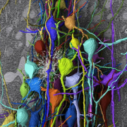
Serial section electron microscopy (ssEM), a technique where volumes of tissue can be anatomically reconstructed by imaging consecutive tissue slices, has proven to be a powerful tool for the investigation of brain anatomy. Between the process of cutting the slices—or “sections”—and imaging them, however, handling 100-106 delicate sections remains a bottleneck in ssEM, especially for batches in the “mesoscale” regime, i.e.,102-103 sections. Our lab is developing a tissue section handling device that transports and positions sections accurately and repeatably for automated, robotic section pick-up and placement onto an imaging substrate.
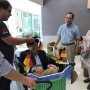
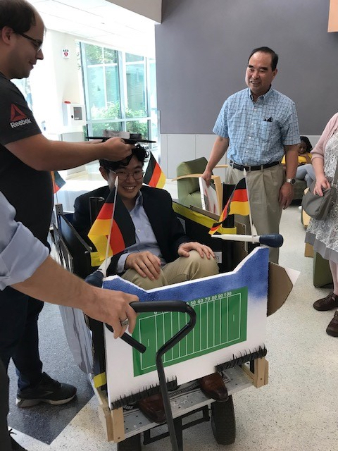 Dr. Timothy Lee successfully defended his thesis this summer. His thesis focused on automating the collection of serial nano-sections sections used to image and analyze up to 1 cubic milimieter of tissue!