
Neuroanatomical Imaging
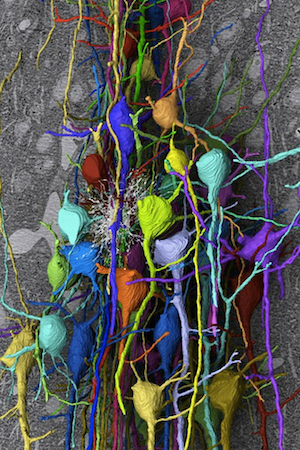
Image courtesy of Sebastian Seung
Serial section electron microscopy (ssEM), a technique where volumes of tissue can be anatomically reconstructed by imaging consecutive tissue slices, has proven to be a powerful tool for the investigation of brain anatomy. Between the process of cutting the slices—or “sections”—and imaging them, however, handling 100-106 delicate sections remains a bottleneck in ssEM, especially for batches in the “mesoscale” regime, i.e.,102-103 sections. Our lab is developing a tissue section handling device that transports and positions sections accurately and repeatably for automated, robotic section pick-up and placement onto an imaging substrate.
Selected Publications
T. Lee, Batch processing of brain tissue sections for millimeter-scale serial section transmission electron microscopy connectomics, Doctoral Dissertation, Summer 2019
[PDF]Timothy J. Lee, Mighten C. Yip, Aditi Kumar, Colby F. Lewallen, Daniel J. Bumbarger, R. Clay Reid and Craig R. Forest, Capillary-Based and Stokes-Based Trapping of Serial Sections for Scalable 3D-EM Connectomics, eNeuro 24 February 2020, 7 (2) ENEURO.0328-19.2019; DOI: https://doi.org/10.1523/ENEURO.0328-19.2019
[PDF]T.J. Lee, A. Kumar, A.H. Balwani, D. Brittain, S. Kinn, C.A. Tovey, E.L. Dyer, N.M. da Costa, R.C. Reid, C.R. Forest*, D.J. Bumbarger* (*co-corresponding authors). Large-scale neuroanatomy using LASSO: Loop-based Automated Serial Sectioning Operation, PLOS One 13(10): e0206172. https://doi.org/10.1371/journal.pone.0206172 (2018)
[PDF]T.J. Lee, C.F. Lewallen, D.J. Bumbarger, P.J. Yunker, R.C. Reid, C.R. Forest. Transport and trapping of nanosheets via hydrodynamic forces and curvature-induced capillary quadrupolar interactions, Journal of Colloid and Interface Science (2018). DOI: 10.1016/j.jcis.2018.07.068
[PDF]J. Lee, I. Kolb, C.R. Forest, C.J. Rozell, Cell membrane tracking in living brain tissue using differential interference contrast microscopy, IEEE Transactions on image processing, 2018 Apr; 27(4):1847-1861. doi: 10.1109/TIP.2017.2787625.
A.S. Chuong, M.L. Miri, L.C. Acker, S.B. Kodandaramaiah, M.A. Henninger, M. Ogawa, R.C. Bandler, N.C. Klapoetke, X. Gu, B.D. Allen, C.R. Forest, B.Y. Chow, X. Han, J.A. Cardin, E.S. Boyden, Noninvasive optical inhibition with a red-shifted microbial rhodopsin, Nature Neuroscience. Vol 17, p. 1123-1129, July 2014.